Overview
Atomic force microscopy, neutron scattering and computer simulations are the three major techniques in use in the NanoBioPhysics Lab along with a several cell biology approaches. A number of complementary techniques are also in use, including: static and dynamic light scattering, calorimetry, optical and electron microscopies, Raman and infrared spectroscopies and a palette of different biochemical tools.
The NanoBioPhysics Lab's Bio-Atomic Force Microscope
The Lab runs a brand-new new-generation bio-Atomic Force Microscope (DriveAFM, Nanosurf) mounted on an inverted fully-motorised optical epi-fluorescence microscope (Axio Observer 7, Zeiss) for correlative studies. The system is a tip-scanning AFM capable of both piezo and photothermal actuation of the cantilever. It is equipped with a number of accessories and software packages that expand its capability including: Petri-dish holder, 150 micro-meter z-stage, Cooling & heating sample holder, Environmental control chamber, Temperature & CO2 controllers, Variable magnetic field generator, Fast scan, FluidFM, Advance force spectroscopy software package, nanorheology software package.
Other AFM systems accessible to us
We have also access to a wide range of commercial AFMs that can achieve sub-nanometer vertical resolution under liquid conditions and can be combined with various extension kits allowing temperature control and in situ fluid exchange. These include: 3 Asylum Research MFP-3D AFMs, 2 Bruker Dimension ICON AFMs, and an Asylum Research Cypher AFM.
Neutron scattering
Neutron scattering is used to probe both the structure and dynamics of the systems of interest as, for example, lipid vesicles or hydrated proteins. Among neutron scattering approaches, we currently use neutron reflectivity, small-angle neutron scattering, neutron spin-echo, and elastic & quasi-elastic neutron scattering. The neutron scattering investigations are carried out at some of the largest neutron scattering facilities worldwide, including the National Institute of Standards & Technology (USA), the Oak-Ridge National Laboratory (USA), the Paul Scherrer Institute (Switzerland), the Institute Laue-Langevin (France), the Rutherford Appleton Laboratory (UK), the Heinz-Maier-Leibnitz Zentrum (Germany), and the Australian Centre for Neutron Scattering (Australia).
Computer simulations:
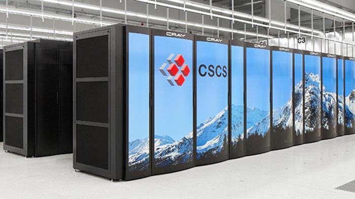
Classical atomistic molecular dynamics (MD) simulations and ab-initio computations are routinely employed in our investigations via the use of well-established packages like Gromacs and CPMD running on supercomputers in a number of different high-computing centres worldwide like ICHEC (IE) and CSCS (CH) pictured, respectively, on the left and right figures below. Recently, we used MD simulations to study the interaction between two phosphonium-based ionic liquids, a mono- and di-cation, with a phospholipid bilayer, with the aim to shed a light on the marked antibacterial effect of the di-cation in comparison to the mono-cation.
Classical atomistic molecular dynamics (MD) simulations and ab-initio computations are routinely employed in our investigations via the use of well-established packages like Gromacs and CPMD running on supercomputers in a number of different high-computing centres worldwide like ICHEC (IE) and CSCS (CH) pictured, respectively, on the left and right figures below. Recently, we used MD simulations to study the interaction between two phosphonium-based ionic liquids, a mono- and di-cation, with a phospholipid bilayer, with the aim to shed a light on the marked antibacterial effect of the di-cation in comparison to the mono-cation.

Cell biology approaches:
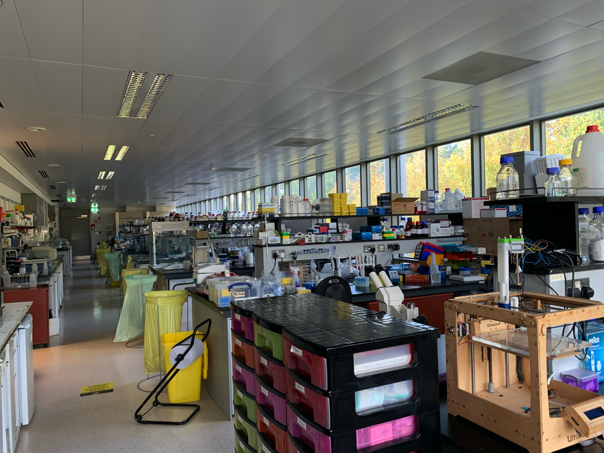
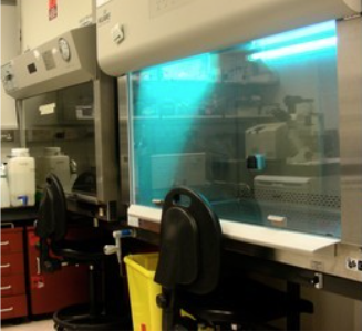
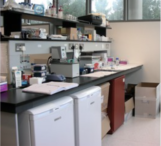
Several cell biology approaches are routinely used in our investigations including, for example, cell survival assays (e.g., MTT), flow cytometry, western blotting of key/targeted proteins, cell migration, colony scattering, and cell invasion assays. We have access to a fully-equipped wet lab facility for biological sample preparation including, fume hoods, centrifuges, balances, temperature controlled storage areas etc. We have also access to a shared tissue culture facility that houses all equipment needed for the growth, maintenance, and analysis of cells. Laminar flow hoods for the sterile handling of cells, as well as several atmosphere-controlled incubators for growing cells are available. Additional phase-contrast, fluorescence and standard bright-field microscopes are accessible for cell analysis. A highly purified water supply, fridges, freezer and cryopreservation system are available for media preparation and storage.
Several cell biology approaches are routinely used in our investigations including, for example, cell survival assays (e.g., MTT), flow cytometry, western blotting of key/targeted proteins, cell migration, colony scattering, and cell invasion assays. We have access to a fully-equipped wet lab facility for biological sample preparation including, fume hoods, centrifuges, balances, temperature controlled storage areas etc. We have also access to a shared tissue culture facility that houses all equipment needed for the growth, maintenance, and analysis of cells. Laminar flow hoods for the sterile handling of cells, as well as several atmosphere-controlled incubators for growing cells are available. Additional phase-contrast, fluorescence and standard bright-field microscopes are accessible for cell analysis. A highly purified water supply, fridges, freezer and cryopreservation system are available for media preparation and storage.
Complementary approaches:
A series of complementary approaches are also available locally to us, which include static and dynamic light scattering, calorimetry, optical and electron microscopies, Raman and infrared spectroscopies and a palette of different biochemical methods.
A series of complementary approaches are also available locally to us, which include static and dynamic light scattering, calorimetry, optical and electron microscopies, Raman and infrared spectroscopies and a palette of different biochemical methods.